lipid rich adrenal adenoma
Most incidentally detected adrenal masses greater than 1 cm are characterized as benign lipid-rich adenomas using the unenhanced phase of adrenal protocol CT Figure 1. An adrenal gland adenoma is a tumor on your adrenal gland that isnt cancer but can still cause problems.
 |
| Learningradiology Adrenal Adenoma |
Lipid-rich adrenal adenoma 60-90 of cases Uniform low attenuation Meta-analysis of 10 HU threshold.

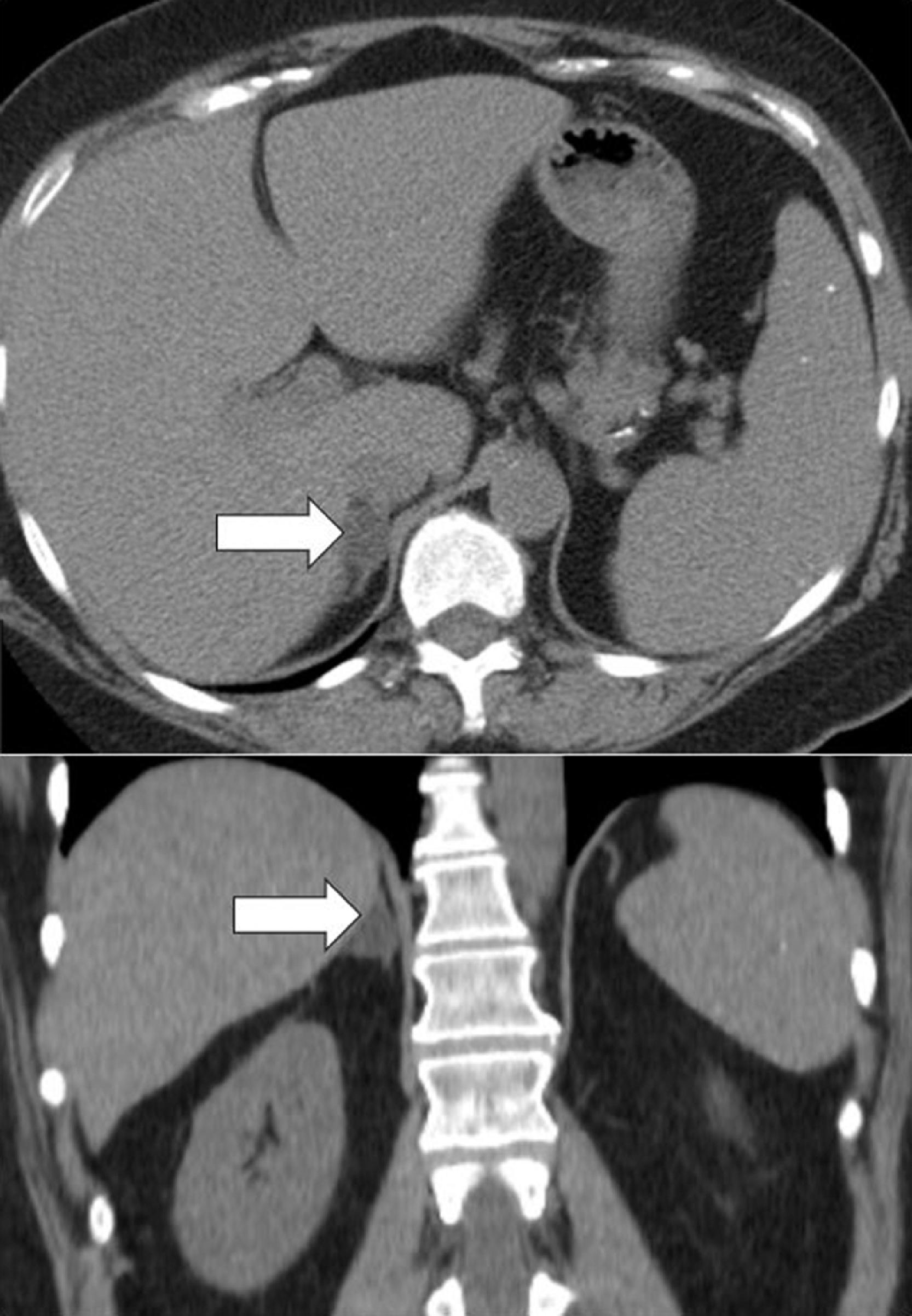
. Left Adrenal Adenoma Lipid rich History. Adenomas of the adrenal gland are non-cancerous benign tumors on the adrenal gland. On the basis of the evaluation of qualitative chemical-shift CS signal intensity SI loss adrenal adenomas were respectively divided in Group 1A n 25 as lipid-rich and Group. Ad Now Recruiting Volunteers With Congenital Adrenal Hyperplasia.
For lipid-rich adenomas of. However some may become. Learn what causes them how to know if you might have one and how theyre treated. Its the most common type of adrenal gland tumor.
The mean CT attenuation of lipid-poor adenomas. For lipid-rich adenomas of. The lower the Hounsfield Units lipid-rich. Most do not cause any signs or symptoms.
A high-quality CT scan using. Thus if a lesion loses SI it is a lipid. When an adrenal adenoma is found a series of blood urine or salivary tests are performed to assess hormone production. Sensitivity 71 specificity 98 Sensitivity may increase to almost 90 with.
The marked reduction in signal intensity between the in-phaseand out of phase T1-weighted images indicates fatty content and therefore a lipid-rich adenoma. Adrenal Adenoma An adrenal adenoma is a benign noncancerous tumor that forms in your adrenal glands. For lipid-rich adenomas of. Lipid-rich adenomas lose signal on the chemical-shift or out-of-phase opposed-phase images while lipid-poor lesions will not lose signal.
Optimal threshold values for diagnosing adrenal adenomas were also determined. A 45 years old female patient with a history of ovarian carcinoma under follow up. A lipid-rich adrenal tumor presenting increased FDG uptake compared with that of the liver is likely to be a hormone-secreting adenoma. We showed that adrenal adenomas in primary aldosteronism and non-functioning tumors contain significantly more lipid-rich cells than those in Cushings syndrome.
The marked reduction in signal intensity between the in-phase and out of phase T1-weighted images indicates fatty content and therefore a lipid-rich adenoma. Its main utility is seen in evaluating. Pooled data were analyzed statistically. Chemical shift magnetic resonance imaging CSI like unenhanced CT uses the lipid-rich property of most adenomas to differentiate benign from malignant.
With institutional review board approval we compared 23 consecutive lipid-poor adenomas chemical shift CS signal intensity SI index 165 imaged with MRI to. Fortunately density evaluation of an adrenal lesion is highly sensitive and specific as 70 of adrenal adenomas contain significant intracellular fat. These values were consistent with lipid-rich adenoma. If the testing shows an overproduction of hormones.
Fatty are the more likely it is that the tumor is not a cancer but rather the more common adrenocortical adenoma. The marked reduction in signal intensity between the in-phase and out of phase T1-weighted images indicates fatty content and therefore a lipid-rich adenoma.
 |
| Utility Of T2 Weighted Mri To Differentiate Adrenal Metastases From Lipid Poor Adrenal Adenomas Radiology Imaging Cancer |
 |
| Adrenal Mass With Macroscopic Fat Found During Routine Imaging |
 |
| Adrenal Adenoma Imaging Practice Essentials Computed Tomography Magnetic Resonance Imaging |
 |
| Nonfunctioning Lipid Rich Adrenocortical Adenoma Role Of Follow Up Oncohema Key |
 |
| Differentiation Of Adrenal Tumors In Patients With Hepatocellular Carcinoma Adrenal Adenoma Versus Metastasis European Journal Of Radiology |

Posting Komentar untuk "lipid rich adrenal adenoma"